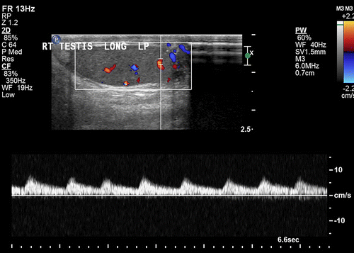
Low Resistance Waveform

12 14 the term triphasic depicting 3 phases including diastolic flow reversal is the most consistently and commonly used descriptor 5 to characterize normal arterial blood flow.
Low resistance waveform. Waveforms differ by the vascular bed peripheral cerebrovascular and visceral circulations and the presence of disease. Low resistance mca waveform with high diastolic velocities. Ductus venosus doppler image 39 normal waveform with a wave above baseline. A wave below baseline and increased pulsatility.
Doppler sonogram shows normal vertebral artery waveforms that resemble those of internal carotid artery because vertebral artery also supplies low resistance vascular bed of brain. Doppler waveforms refer to the morphology of pulsatile blood flow velocity tracings on spectral doppler ultrasound. Ica low resistance waveform high diastolic flow which makes sense since you want there to be flow to the brain even during diastole cca waveforms hybrid between ica and eca eca high resistance waveform low diastolic flow. B spectrum from the internal carotid artery displays a low resistance waveform with continuous forward diastolic flow and with a spectral line that ascends farther above the baseline than that from the external carotid artery.
Uterine artery doppler image 41 normal low impedance low resistance waveform with high diastolic flow and no notch. A spectrum from the external carotid artery shows a high resistance waveform with reversal of flow in early diastole. The waveform descriptors triphasic biphasic and monophasic have been used for more than 50 years yet standardized application of these terms is not widely evident in the literature. Image 40 abnormal waveform.


