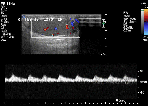
High Resistance Waveform

Normal flow velocities for adult common femoral superficial femoral popliteal and tibioperoneal arteries are in the range of 100 cm sec 80 90 cm sec 70 cm sec and 40 50 cm sec respectively 6.
High resistance waveform. There is no antegrade flow at end diastole end diastolic velocity is 0. There is coexisting pulsatility of venous waveforms and often a soft tissue bruit which is the color speckling at the site of the fistula caused by tissue reverberation from the adjacent highly turbulent flow. 12 14 the term triphasic depicting 3 phases including diastolic flow reversal is the most consistently and commonly used descriptor 5 to characterize normal arterial blood flow. Doppler waveforms refer to the morphology of pulsatile blood flow velocity tracings on spectral doppler ultrasound.
Waveforms differ by the vascular bed peripheral cerebrovascular and visceral circulations and the presence of disease. Although this is a more typical doppler waveform of an artery supplying a high resistance vascular bed such as the resting limbs it is a pathologic finding in the evaluation of the va and may indicate cephalad vertebrobasilar occlusive disease. These pulsatile waveforms with the end diastolic velocities within 20 25 of peak systolic values indicate high resistance to arterial flow. Triphasic high resistance waveforms are seen in lower limb arteries as in other peripheral arteries fig 9.
However triphasic has also.


